![]()
Designed for Reconstructive Plastic Surgery, the Exashape membrane is processed through a proprietary method that maintains the unique qualities of natural bovine pericardium¹.
Exashape Bioshield pocket guarantees fast integration times with cell ingrowth², minimal inflammation³ and negligible serum production, while maintaining remarkable mechanical properties¹.
Our ultra light bovine membrane has two different layers
COMPACT LAYER
Provides structural support, reactivating fibroblasts and VEGF2 which enable the reparative process with formation of new tissue and blood vessels.
FIBROUS LAYER
Highly porous, allows cytokines and growth factors to trigger the process of immediate revitalisation of endogenous connective tissue and early neoangiogenesis with high neoformation of vessels.
The bilayer membrane undergoes a 3-stage process of revitalization by endogenous connective tissue.
During implantation, the collagen matrix absorbs the blood and the revitalization process begins immediately, characterized by a controlled and short-lived inflammation phase with considerable neoformation of vessels [2,3].
Fibroblasts reactivate collagen by triggering the ingrowth of new vessels, which provide the metabolic needs. At this stage of the reparative process, cell proliferation prevails, which targets the growth of new tissue [2].
The actual remodeling phase begins its course and is the result of the precise balance in the synthesis of collagen, which becomes an integral part of the tissue [3].
The cell-friendly process preserves the active elements in the repair process within the membrane: proteoglycans, hyaluronic acid, fibronectin, elastin and of course, native collagen. They represent a natural reservoir of bioactive factors, which participate in the revitalization process by controlling inflammation while promoting cell proliferation and migration [2]. We limit the amount of biological mass implanted (up to 50% less, due to lower thickness of pericardium compared to dermis) promoting integration even in cases of poor blood supply, while maintaining the highest bio-mechanical performance [1,4].
![]()
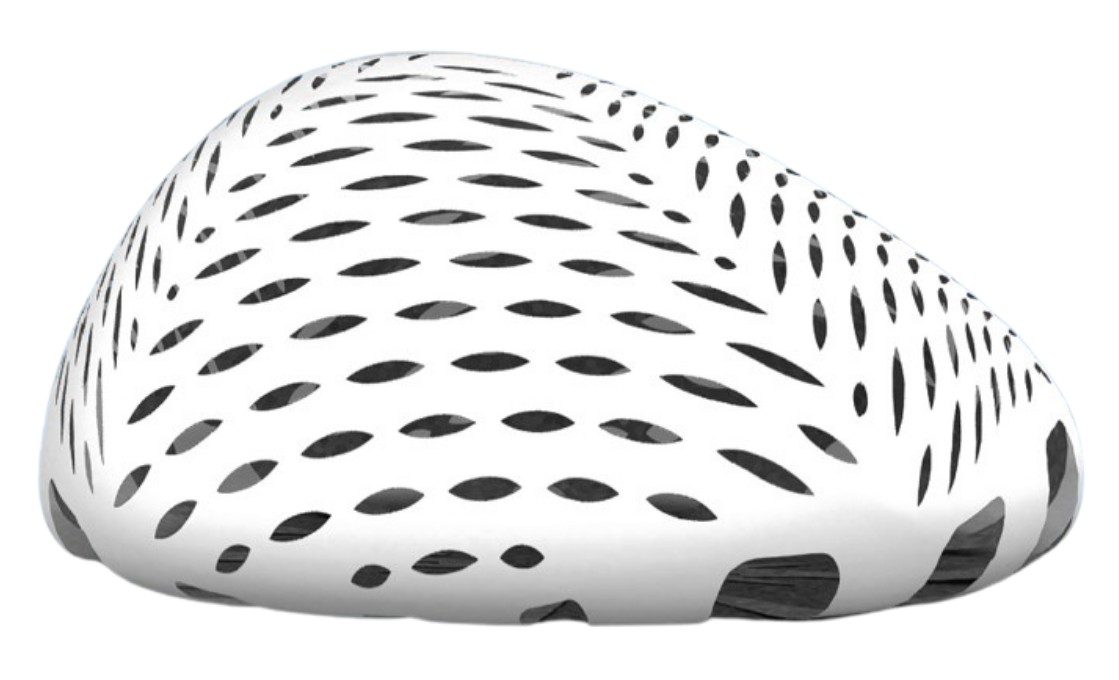
The membrane’s shape is conceived for anatomical and round implants. A perfect support is achieved by tightening the petals so that wrinkle-free tailor-made protection for each implant profile or volume, without waving or wrinkling which, as with thick matrices, can be sometimes perceptible in thinner patients.
![]()
Less than 0.6 mm thick, this membrane helps to minimize the foreign body response by facilitating rapid integration and faster tissue regeneration. The overall biological mass is 50% less than other membranes dermal based.
![]()
The meshed scaffold design optimizes fluid drainage preventing accumulation. The perfect balance in the proportion between cut-outs and collagen best support timing of regeneration.
![]()
Bioshield pocket 3D adapts more effectively to the implant and it shields only where needed: the dermal flap interface. This way foreign body reaction is minimaized, and also healing process is not overloaded with unnecessary effort to remodeling exceeding biomass.
The new pre-shape design facilitates a rapid, touchless assembly procedure that can be completed in less than 1 minute minimizing the risk of contamination and ensuring a sterile environment far optimal surgical outcomes.
PREPECTORAL
(W x H x Proj.)
| Code | Model | Size |
|---|---|---|
| AEPB(F)154-158H050 | SIZE A | 15,5 x 15 x 5 cm |
| AEPB(F)174-178H055 | SIZE B | 17,5 x 17 x 5,5 cm |
| AEPB(F)194-198H065 | SIZE C | 19,5 x 19 x 6,5 cm |
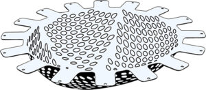
A patented design mesh that improves surgical procedures.
Bioshield Pocket® serving as an elective option for immediate PrePectoral breast reconstruction for the protection of the implant/tissue interface
Bioshield Pocket® aims to streamline surgical procedure as the rehydrated membrane allows for quick wrapping of the implant without the need for complex tailoring procedures, reducing intra-operative delays, lowering risks of implant damage and potential contamination

After running the threads through the “petal” holes, the mesh is rehydrated in the sterile tray, with the smooth side facing the operator. Subsequently, the anterior surface of the implant is positioned on the membrane within a few seconds. The ‘petals’ are tightened to the implant’s posterior side creating a “purse string” assembly.
Due to the meshed pattern and rough external surface, Bioshield Pocket® offers a secure grip on the chest wall ensuring that the implant remains in place during and after surgery.
The high drapability allows the membrane to follow the silicone implant shape with no “dead spaces” or wrinkles
PREPECTORAL

(W x H x TH)
| Code | Model | Size |
|---|---|---|
| AEPB(F)144-188S | SMALL | 18 x 14 cm x 0,5 mm |
| AEPB(F)164-208S | MEDIUM | 20 x 16 cm x 0,5 mm |
| AEPB(F)184-228S | LARGE | 22 x 18 cm x 0,5 mm |
| AEPB(F)204-238S | EXTRA LARGE | 23 x 20 cm x 0,5 mm |
Including 2 X resorbable monofilament PGCL sutures for the preparation of the “purse string” assembly

| Code | Model | Size |
|---|---|---|
| POCKET S 4.0 | KIT SMALL | 18 x 14 cm x 0,5 mm |
| POCKET M 4.0 | KIT MEDIUM | 20 x 16 cm x 0,5 mm |
| POCKET L 4.0 | KIT LARGE | 22 x 18 cm x 0,5 mm |
| POCKET XL 4.0 | KIT EXTRA LARGE | 23 x 20 cm x 0,5 mm |
Exashape Prepec is a rectangular meshed membrane. Thanks to its expansion ratio, after rapid rehydration it is able to cover implants of all shapes and sizes, with minimum handling and maximum stretchability/drapability

PREPECTORAL


(Wx H x TH)
| Code | Model | Size |
|---|---|---|
| AEPB(F)058-100S | Rectangular | 10 x 5 cm x 0,5 mm |
| AEPB(F)058-200S | Rectangular | 20 x 16 cm x 0,5 mm |
| AEPB(F)108-150S | Rectangular | 15 x 10 cm x 0,5 mm |
| AEPB(F)108-200S | Rectangular | 20 x 10 x cm x 0,5 mm |
| AEPB(F)158-200S | Rectangular | 20 x 15 cm x 0,5 mm |
| AEPB(F)158-150S | Squared | 15 x 15 cm x 0,5 mm |
exaShape Grid comes in 2 shapes:
3 x Half Moon
2 x Rectangular

They are both fenestrated with circular holes to optimize fluid drainage capacity. The membrane rehydrates quickly and is immediately available for use.
The range of sizes allows its immediate use in most patients
SUBPECTORAL


(W x H x TH)
| Code | Model | Size |
|---|---|---|
| AEPB(F)084-162S | Half Moon | 10 x 5 cm x 0,5 mm |
| AEPB(F)074-172S | Half Moon | 20 x 5 cm x 0,5 mm |
| AEPB(F)104-202S | Half Moon | 15 x 10 cm x 0,5 mm |
| AEPB(F)104-150S | Rectangular | 15 x 15 cm x 0,5 mm |
| AEPB(F)104-200S | Rectangular | 20 x 15 cm x 0,5 mm |
Exashape Expander is used in combination with a breast expander in breast reconstruction procedures as a means of pocket closure.
It is ‘circular-hole’ fenestrated to optimize drainage capacity of fluids

SUBPECTORAL

(W x H x TH)
| Code | Model | Size |
|---|---|---|
| AEPB(F)064-080S | Rectangular | 8 x 6 cm x 0,5 mm |
Exashape Nac® is a perforated membrane used in breast reconstruction procedures for the areola-nipple area in order to create supplementary tissue, to cover a nipple reconstruction device or for a stand-alone use. It is fenestrated with circular holes to optimise fluid drainage capacity

(W x H x TH)
| Code | Model | Size |
|---|---|---|
| AEPB(F)084-080S | Round | 8 x 8 cm x 0,5 mm |
PROTECTION OF
UPPER QUADRANTS
PROTECTION OF
LOWER QUADRANTS
IN CONJUNCTION WITH
BIOSHIELD POCKET

In addition, it can be used for wrapping a breast implant in combination with the Bioshield Pocket® membrane to achieve greater coverage, increasing containment capacity, and allowing the coverage of larger implants

(W x H x TH)
| Code | Model | Size |
|---|---|---|
| AEPB(F)080-099S | SIZE A | 8 x 9 cm x 0,5 mm |
| AEPB(F)100-119S | SIZE B | 10 x 11 cm x 0,5 mm |
| AEPB(F)120-139S | SIZE C | 12 x 13 cm x 0,5 mm |
| AEPB(F)140-159S | SIZE D | 14 x 15 cm x 0,5 mm |
| AEPB(F)160-179S | SIZE E | 16 x 17 cm x 0,5 mm |

![]()
![]()
REFERENCES